CURRENT STATUS OF ULTRASOUND DIAGNOSING FOR PREGNANT BOTTLENOSE DOLPHINS (TURSIOPS TRUNCATUS) IN ENOSHIMA AQUARIUM
2013年09月
41st IMATA Conference 2013, Las Vegas (Poster Presentation)
PREGNANT BOTTLENOSE DOLPHINS (TURSIOPS TRUNCATUS)
IN ENOSHIMA AQUARIUM
Fumio Terasawa(1),Hiroshi Koie(2),Hiroki Chino(3),Yukiko Hori(1)
(1) Enoshima Aquarium, Enoshima, Japan
(2) Nihon University,
College of Bioresource Sciences Veterinary Physiology, Fugisawa, Japan
(3) Sonic Japan K.K, Tokyo, Japan
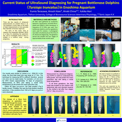
In the late 1980s, we learned husbandry behaviors from IMATA, and the first voluntary blood sampling was started with Pacific white-side dolphins(Lagenorhynchus obliquidens) in 1990. After that, we observed several physiological changes during pregnancy with rectal temperatures (Terasawa, Yokoyama, & Kitamura, 1999) and blood data (Terasawa, & Kitamura, 2005; Terasawa, Arai, Tokura, & Ohshita, 2008). In Japan, ultrasound has been used as one way to diagnose early pregnancy of cetaceans, as well as in North America. In Enoshima Aquarium, the first ultrasound diagnosing for a pregnant dolphin was a finless porpoise (Neophocaena asiaeorientalis) in a drained pool in 1992. Currently we perform ultrasound diagnosing to investigate fetus growth and to predict the exact parturition day.
From 2005 to 2013, ultrasound diagnosing by husbandry has been performed for five pregnancies in four Pacific bottlenose dolphins(Tursiops truncatus gilli). In three of these cases, the number of observations ranged between 30 to 60 times during the whole pregnancy. Examinations were performed with a Sono Site Micro MAXX and C60e/5-2MHz transducer. A frequency of 3.5MHz was used. Additionally, a photograph was taken simultaneously at the time of ultrasound diagnosing in order to investigate where a fetus was located in a mother’s body.
On 30 June 2013, two dolphins were pregnant and the ultrasound examination was not been finished. At present, 74 measurements of the bioparietal diameter of the fetus have been gathered and more data will be measured. There is a significant relationship between the bioparietal diameters and the days before parturitions (p<0.001). In this abstract, however, we are not able to show the results. The earliest data was obtained at 326 days pre-partum, while the latest data was at 4 days pre-partum. The head of the fetus was located in the mother’s left side abdomen toward to mother’s tail flukes at 4 days pre-partum.
On 1 June 2012, a female bottlenose dolphin was born in Enoshima Aquarium. Her birth was only one day later than the parturition day predicted by ultrasound diagnosing. As a result of analyzed blood taken from the umbilical cord, it became clear that she was a 5th generation dolphin in captivity. The present study aims not only investigate fetus growth by ultrasound diagnosing, but also to collect enough data needed to confirm this statistically.
This work was supported by the Cooperation Research Program of Wildlife Research Center, Kyoto University.
References
Terasawa, F., Yokoyama, Y., & Kitamura, M. (1999). Changing the rectal temperature at a parturition in bottlenose dolphins. Zoo Biology, 18, 153-156.
Terasawa, F. & Kitamura, M. (2005). Hyperlipemia of captive bottlenose dolphins during Pregnancy. Journal of Veterinary Medical Science, 6, 341-344.
Terasawa, F., Arai, T., Tokura, T., & Ohshita, I. (2008). Changes in fibrinogen concentrations in captive bottlenose dolphins during pregnancy. Journal of Veterinary Medical Science, 70, 1277-1279.
★2nd Place of Poster Presentation★